OFA Testing

OFA testing
The Orthopedic Foundation for Animals (OFA) was founded as a private, not-for-profit foundation whose main goal is to improve the health and well-being of companion animals through a reduction in the incidence of genetic disease via OFA testing.
OFA testing databases
The OFA databases are central to the organization’s objective of establishing control programs to lower the incidence of inherited disease. They serve all breeds of dogs and cats, provide veterinarians with broad ranged medical screening, and provide breeders with a means to respond to the challenge of improving the genetic health of their breed through better breeding practices
We offer OFA (Orthopedic Foundation for Animals) screening for the following:
- Basic Cardiac Evaluations by Practitioner
- Patella Evaluations by Practitioner
- Radiology Imaging and Submission for Hip Evaluation
- Radiology Imaging and Submission for Elbow Evaluation

Requirements for Radiology Submissions
- Preliminary Evaluations- for breeders with dogs 4 to 23 months old to make placement decisions
- Full Evaluations- Dogs must be over 2 years of age
- Your pet’s Registered name, call name, date of birth, breed, microchip number, and parentage information is required to be labeled on the radiograph films. Please make sure to fill all of this information out on your OFA testing application form prior to your appointment. These forms can be found here: https://ofa.org/applications/
Please note
For accurate positioning and diagnosis, your pet will be given chemical sedation appropriate for his/ her size and energy level. This may include oral or injectable versions.
*We will not attempt to perform these radiographs on an animal that is hyper, stressed, aggressive, and/or fearful without appropriate medication(s).*
The veterinarian overseeing your pet’s case will advise and communicate with you about the care and medication needs of your animal for this service.
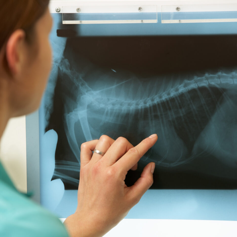
These radiographs are taken in our clinic and then are submitted to the OFA to be evaluated by a radiologist for hip/ elbow congruity, subluxation, the condition of the acetabulum, and the size, shape, and architecture of the femoral head and femoral neck.
Radiographs submitted to the OFA should follow the American Veterinary Medical Association recommendations for positioning. This view is accepted world wide for detection and assessment of hip joint irregularities and secondary arthritic hip joint changes. To obtain this view, the animal must be placed on its back in dorsal recumbency with the rear limbs extended and parallel to each other. The knees (stifles) are rotated internally, and the pelvis is symmetric. Chemical restraint to the point of relaxation is recommended.
For elbows, the animal is placed on its side and the respective elbow is placed in an extreme flexed position.
The radiograph must be permanently identified with the animal’s registration number or name, the date the radiograph was taken, and the veterinarian’s name or hospital name. If this required information is illegible or missing, the OFA cannot accept the film for registration purposes. Both the owner and vet should complete and sign their respective sections of the OFA testing application. It is important to record on the OFA application the animal’s tattoo or microchip number in order for the OFA to submit results to the AKC. Sire and dam information should also be present.
Hip Radiology of females in estrus or pregnant should be avoided due to possible increased joint laxity (subluxation) from hormonal variations. OFA testing recommends radiographs be taken one month after weaning pups and one month before or after a heat cycle. Physical inactivity because of illness, weather, or the owner’s management practices may also result in some degree of joint laxity. The OFA recommends evaluation when the dog is in good physical condition.
Three different radiologists review the radiographs and a score is given based on the dog’s hips/ elbow conformation relative to other dogs of the same age and breed.
The OFA classifies hips into seven different categories: Excellent, Good, Fair (all within Normal limits), Borderline, and then Mild, Moderate, or Severe (the last three considered Dysplastic).
- Excellent: Superior conformation; there is a deep-seated ball (femoral head) that fits tightly into a well-formed socket(acetabulum) with minimal joint space.
- Good: Slightly less than superior but a well-formed congruent hip joint is visualized. The ball fits well into the socket and good coverage is present.
- Fair: Minor irregularities; the hip joint is wider than a good hip. The ball slips slightly out of the socket. The socket may also appear slightly shallow.
- Borderline: Not clear. Usually, more incongruency present than what occurs in a fair but there are no arthritic changes present that definitively diagnose the hip joint being dysplastic.
- Mild: Significant subluxation present where the ball is partially out of the socket causing an increased joint space. The socket is usually shallow, only partially covering the ball.
- Moderate: The ball is barely seated into a shallow socket. There are secondary arthritic bone changes usually along the femoral neck and head (remodeling), acetabular rim changes (osteophytes or bone spurs) and various degrees of trabecular bone pattern changes
(sclerosis). - Severe: Marked evidence that hip dysplasia exists. The ball is partly or completely out of a shallow socket. Significant arthritic bone changes along the femoral neck and head and acetabular rim changes.
Once submitted, you should expect to receive your OFA screening certificate from the OFA radiologists within 3-4 weeks.
For more OFA testing information, please visit:
Services
We are committed to providing compassionate primary pet care for your pet, treating both of you with dignity and respect. Above all, our goal is to enhance the human-animal bond and ensure it thrives. Consequently, as Richmond and surrounding communities grow, we plan to expand our practice, adding skilled doctors and team members to meet increasing needs.

